Drag to orbit · Scroll or pinch to zoom Shift-drag to pan · Keyboard: arrows orbit, + / − zoom, R resets
Matching cut surfaces
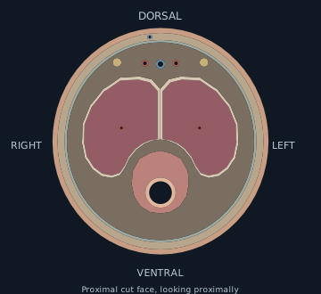
Same tissue, opposite viewing directions
Proximal stump Looking proximally · R at leftRIGHT Dorsal ↑ · Ventral ↓ LEFTAmputated segment Looking distally · L at leftLEFT Dorsal ↑ · Ventral ↓ RIGHT
Surface shapes come from triangle-plane intersections. Small vessel walls and nerves are schematic. Select a structure in either section.
Supplementary repair close-ups
Inspect an individual repair.
The full movie above repairs the original amputated anatomy. Use these separate detail models to inspect the wall, lumen, and device mechanism.
Teaching order, not a prescribed operative order. These are constructed, direct repairs with matching ends and no modeled tissue loss.
Enlarged schematic · display proportionsDrag to orbit · Pinch to zoom Arrows orbit · + / − zoom · R restores viewTunical closureUrethral wallArteryVeinNerve + wrap
DVRLProxDist
Repair sequence
The 3D repair viewer could not start. The teaching text and sources remain available.
Corpora: tunical edges close while the attached urethra, vessels, and nerves remain divided.
Urethra: matching wall edges meet around a catheter, preserving the channel.
Microvascular: an artery joins its arterial partner; a vein joins its venous partner. Each repair engages the walls without crossing the lumen.
Nerve: a schematic Nerve Tape wrap surrounds aligned, filled nerve ends. The short leaf closes before the long overlapping leaf. Reviewed evidence does not establish penile replantation outcomes.
Awaiting repair:UrethraArteriesNervesCut ends stay separated during tunical closure.
IdentifyApproximateSutureInspect
Identify matching ends
Enlarged sutureMoving thread-path marker
Translucent SIS wrapFilled nerve + epineurium
The moving marker traces a thread path; needle handling and knot tying are omitted. Preparation, debridement, clamps, and grafts are not animated here. Skin and fascia are hidden throughout these close-ups; the urethral view also hides surrounding spongiosum. Sutures and wall thickness are enlarged for visibility. The cutaway hides a window or wedge of tissue and its stitches for inspection. An open rendered lumen does not establish perfusion, healing, or clinical patency.
Nerve Tape is a porcine SIS implant with nitinol microhooks. This dorsal nerve scene illustrates the device mechanism; it does not establish a penile indication or predict sensory recovery. The reviewed labeling concerns peripheral nerve discontinuities closed by extremity flexion. Sizing, material sensitivities, preparation, and optional securing sutures require the full manufacturer IFU.
Scope and review status
From anatomy to reconstruction.
One continuous movie follows the original anatomy through amputation, tunical closure, urethral repair, arterial and venous anastomoses, Nerve Tape coaptation, and coverage. The supplementary viewer provides separate detail models for inspection. The storyboard describes the wider reconstructive process. Teaching order does not prescribe operative order.
Other injury patterns
Incomplete injuries retain a tissue bridge and may retain some perfusion. Crush, avulsion, and injuries at other levels change the zone of injury, tissue available for repair, and reconstructive plan. This complete transverse teaching example does not model them.
Questions for anatomical review
Confirm the shaft level, tunical contours, and constructed medial septum.
Confirm the urethral lumen position and the ventral contour of Buck fascia.
Confirm vessel and nerve courses, caliber, and which channels to show as repairable.
Review the illustrative wall bites and the case-specific operative sequence.
Evidence and provenance
Reused geometry: Open3DAnatomy, with Z-Anatomy and BodyParts3D ancestry. New walls, cut faces, skin, fascia, and small structures are constructed additions. Geometry is CC BY-SA 4.0.